Home
/ Shoulder Muscles Diagram Anterior : Muscles Of The Upper Arm And Shoulder Blade Human Anatomy Kenhub Youtube - The shoulder is the region where the upper limb is attached to the trunk.
Shoulder Muscles Diagram Anterior : Muscles Of The Upper Arm And Shoulder Blade Human Anatomy Kenhub Youtube - The shoulder is the region where the upper limb is attached to the trunk.
Shoulder Muscles Diagram Anterior : Muscles Of The Upper Arm And Shoulder Blade Human Anatomy Kenhub Youtube - The shoulder is the region where the upper limb is attached to the trunk.. This diagram depicts shoulder muscle diagram. The shoulder is a mobile structure that allows the arm to move freely in all directions. The main shoulder muscles are trapezius, deltoid, pectoralis major and 4 rotator cuff muscles: The bones of the shoulder are: The tendons, which anchor muscle to bone;
The largest of these shoulder muscles is the. Even though anterior deltoid force is relatively high, its ability to abduct the shoulder is low due to a very small. This flexibility is also what makes the shoulder prone to instability and injury. An anterior view of the deep muscles and ligaments of the shoulder download scientific diagram from www.researchgate.net this muscle moves each shoulder joint in four distinct ways as well as keeps the arms attached to the body. The following is an overview of the shoulder muscle anatomy.
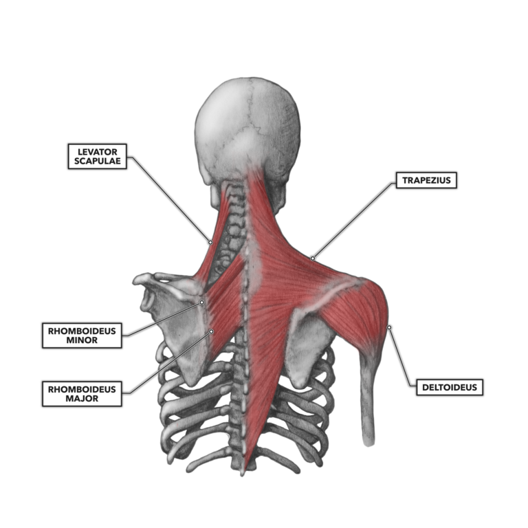
Muscles Of The Shoulder And Arm Dummies from www.dummies.com The main shoulder muscles are trapezius, deltoid, pectoralis major and 4 rotator cuff muscles: Its main function is shoulder flexion, which is characterized by raising your upper arms up to the front and overhead. Located superior to the shoulder joint, the deltoid muscle works with the supraspinatus to abduct the arm at the shoulder. The latissimus dorsi and teres major on the posterior side extend and adduct the arm towards the vertebrae of the back. When autocomplete results are available use up and down arrows to review and enter to select. Shoulder girdle laterally (spine of scapula, acromion, some clavicle) inserts: Muscle anatomy diagram 12 photos of the muscle anatomy diagram canine muscle anatomy diagram, dog muscle anatomy diagram, lower leg muscle anatomy diagram, muscle anatomy of human back, tricep. The shoulder blade (scapula) connects to the collarbone (clavicle) at this joint.
The shoulder blade (scapula) connects to the collarbone (clavicle) at this joint.
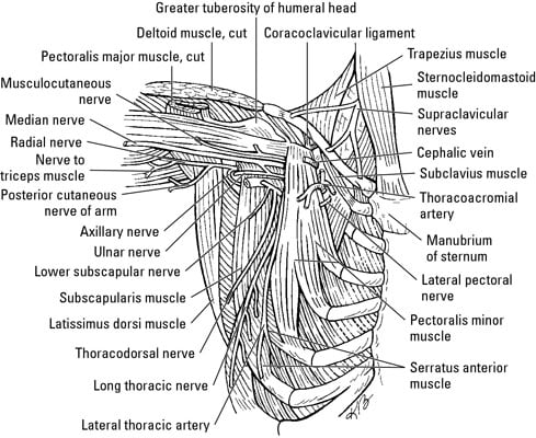
Many people visit the doctor with front, or anterior, shoulder pain. This flexibility is also what makes the shoulder prone to instability and injury. The bursa is a small sac of fluid that cushions and. And the ligaments, which connect bones. Anterior deltoid the anterior deltoid is located on the front of your shoulder. On the anterior side of the shoulder the coracobrachialis serratus anterior pectoralis major and pectoralis minor muscles work as a group to flex and adduct the scapula and humerus anteriorly toward the sternum. This often happens when stress is placed on the tissues that stabilize the shoulder—the muscles; All of them are supplied by the respective branches of the brachial plexus. Anterior shoulder muscles, also called the pectoral muscles, attach the upper extremity to the clavicle and the thoracic cage. These muscles form the outer shape of the shoulder and underarm. Muscle anatomy diagram 12 photos of the muscle anatomy diagram canine muscle anatomy diagram, dog muscle anatomy diagram, lower leg muscle anatomy diagram, muscle anatomy of human back, tricep. The largest of these shoulder muscles is the. What can you tell us about how these joints work?.
Hip muscles anatomy hip anatomy anatomy organs human body anatomy human anatomy and physiology anatomy drawing anatomy male human body muscles muscles of the neck. On the anterior side of the shoulder the coracobrachialis serratus anterior pectoralis major and pectoralis minor muscles work as a group to flex and adduct the scapula and humerus anteriorly toward the sternum. An anterior view of the deep muscles and ligaments of the shoulder download scientific diagram from www.researchgate.net this muscle moves each shoulder joint in four distinct ways as well as keeps the arms attached to the body. The muscle helps to move your scapula in various directions and is essential for proper shoulder function. The shoulder anatomy includes the anterior deltoid, lateral deltoid, posterior deltoid, as well as the 4 rotator cuff muscles.
Crossfit Shoulder Muscles Part 2 Posterior Musculature from www.crossfit.com Its main function is shoulder flexion, which is characterized by raising your upper arms up to the front and overhead. The shoulder muscles are responsible for maintaining the widest range of motion of any joint in your body. Subscapularis, supraspinatus, infraspinatus and teres minor. This often happens when stress is placed on the tissues that stabilize the shoulder—the muscles; Find out in this anatomy of the shoulder quiz. The partner should slowly, but firmly press on both sides of your shoulder to compress the ac joint. The main shoulder muscles are trapezius, deltoid, pectoralis major and 4 rotator cuff muscles: Numerous muscles help stabilize the three joints of.
Shoulder muscles diagram anterior :
And the ligaments, which connect bones. Parts of the right shoulder blade: The most common shoulder injuries are sprains, strains, and tears. Muscle anatomy diagram 12 photos of the muscle anatomy diagram canine muscle anatomy diagram, dog muscle anatomy diagram, lower leg muscle anatomy diagram, muscle anatomy of human back, tricep. The partner should slowly, but firmly press on both sides of your shoulder to compress the ac joint. These muscles form the outer shape of the shoulder and underarm. The infraspinatus muscle is one of the four rotator cuff muscles crossing the shoulder joint and is commonly injured. The shoulder joint is supplied by the anterior and posterior circumflex humeral arteries, which are both shoulder muscles diagram. The shoulder is the region where the upper limb is attached to the trunk. The shoulder is a mobile structure that allows the arm to move freely in all directions. The serratus anterior is a muscle that attaches your shoulder blade, known as your scapula, to your rib cage. Shoulder muscles diagram / anterior and posterior shoulder muscles diagram quizlet : The clavicle (collarbone), the scapula (shoulder blade), and the humerus (upper arm bone) as well as associated muscles, ligaments and tendons.
The bones of the shoulder are: The shoulder is the region where the upper limb is attached to the trunk. The list of muscles and their functions are presented below. These muscles form the outer shape of the shoulder and underarm. Test your knowledge of the clavicle, scapula and humerus with our labeled diagram exercises and quizzes!
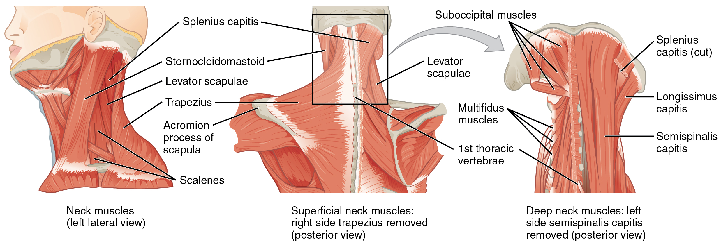
The Ventral Neck Muscles Lecturio Online Medical Library from d3uigcfkiiww0g.cloudfront.net Subscapularis, supraspinatus, infraspinatus and teres minor. The most common shoulder injuries are sprains, strains, and tears. The shoulder joint is supplied by the anterior and posterior circumflex humeral arteries, which are both shoulder muscles diagram. These muscles form the outer shape of the shoulder and underarm. Muscle anatomy diagram 12 photos of the muscle anatomy diagram canine muscle anatomy diagram, dog muscle anatomy diagram, lower leg muscle anatomy diagram, muscle anatomy of human back, tricep. Shoulder muscles diagram / anterior and posterior shoulder muscles diagram quizlet : The bones of the shoulder are: And the ligaments, which connect bones.
The shoulder is not a single joint, but a complex.
The scapula is a triangular shaped bone that functions mainly as a site for muscle attachment. The latissimus dorsi and teres major on the posterior side extend and adduct the arm towards the vertebrae of the back. All of them are supplied by the respective branches of the brachial plexus. The most common shoulder injuries are sprains, strains, and tears. This flexibility is also what makes the shoulder prone to instability and injury. Parts of the right shoulder blade: The shoulder is not a single joint, but a complex. Subscapularis, supraspinatus, infraspinatus and teres minor. Shoulder muscles diagram / anterior and posterior shoulder muscles diagram quizlet : Deltoid tuberosity on humerus action: Shoulder problems may limit arm. On the anterior side of the shoulder the coracobrachialis serratus anterior pectoralis major and pectoralis minor muscles work as a group to flex and adduct the scapula and humerus anteriorly toward the sternum. Anterior shoulder muscles, also called the pectoral muscles, attach the upper extremity to the clavicle and the thoracic cage.
The serratus anterior is a muscle that attaches your shoulder blade, known as your scapula, to your rib cage shoulder muscles diagram. The list of muscles and their functions are presented below.